|
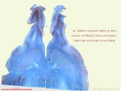
horizontal section of rat forebrain showing architecture of olfactory bulbs. Nissl and myelin stains.
|
| filename, dt_29_NRLFB_1p25
(format, JPEG)
|
| file size, 0.06 MiB |
|
size at 300 dpi, 97mm x 73mm/ 3.84" x 2.88"
|
| date, 20 08 2005
|
| to see the image full-size, click the thumbnail |
|
|
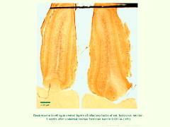
doublecortin labelling of the olfactory bulbs, horizontal section in rat. Note the stained neurons migrating from the rostral migratory streams centrally.
|
| filename, x41DblCrtOBslide
(format, JPEG)
|
| file size, 0.07 MiB |
|
size at 300 dpi, 97mm x 73mm/ 3.84" x 2.88"
|
| date, 31 05 2005
|
| to see the image full-size, click the thumbnail |
|
|
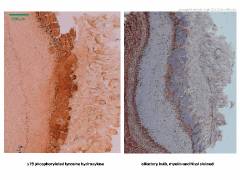
outer layers of the olfactory bulb; left, tyrosine hydroxylase labelling; right, histological stains to demonstrate anatomy
|
| filename, dt_bulb_comparison
(format, JPEG)
|
| file size, 0.08 MiB |
|
size at 300 dpi, 97mm x 73mm/ 3.84" x 2.88"
|
| date, 15 08 2005
|
| to see the image full-size, click the thumbnail |
|
|
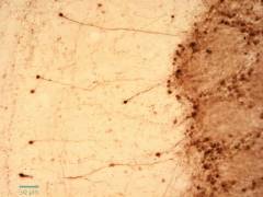
detail of external plexiform (left) and glomerular (right) layers of rat olfactory bulb; tyrosine hydroxylase labelling. Note the periglomerular cells.
|
| filename, dt_29_20x_panTH
(format, JPEG)
|
| file size, 0.06 MiB |
|
size at 300 dpi, 97mm x 73mm/ 3.84" x 2.88"
|
| date, 20 08 2005
|
| to see the image full-size, click the thumbnail |
|
|
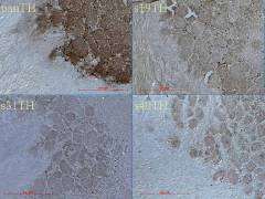
tyrosine hydroxylase phosphorylation variants in the rat olfactory bulb. Rostral is to the left.
|
| filename, dt_olfactory_bulb
(format, JPEG)
|
| file size, 0.06 MiB |
|
size at 300 dpi, 97mm x 73mm/ 3.84" x 2.88"
|
| date, 20 08 2005
|
| to see the image full-size, click the thumbnail |

|
|
olfactory bulb, high power view. Glomerular and nerve cell layers. Labelled for tyrosine hydroxylase phosphorylation variants.
|
| filename, desktopvariantTH
(format, JPEG)
|
| file size, 0.1 MiB |
|
size at 300 dpi, 97mm x 73mm/ 3.84" x 2.88"
|
| date, 20 08 2005
|
| to see the image full-size, click the thumbnail |
|
|
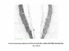
glomerular layers, rat olfactory bulb after 6-OHDA to MFB three weeks previously (left). Tyrosine hydroxylase (s19 phosphorylated) immunohistochemistry.
|
| filename, dt-28_OBx5greyscale
(format, JPEG)
|
| file size, 0.06 MiB |
|
size at 300 dpi, 97mm x 73mm/ 3.84" x 2.88"
|
| date, 23 08 2005
|
| to see the image full-size, click the thumbnail |
|
|
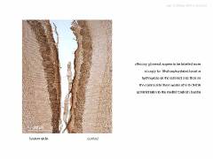
detail of glomeruli of OB after 6-OHDA administration to medial forebrain bundle (left). Tyrosine hydroxylase (s19 phosphorylated) immunohistochemistry.
|
| filename, dt-27_unilat6OHDA
(format, JPEG)
|
| file size, 0.06 MiB |
|
size at 300 dpi, 97mm x 73mm/ 3.84" x 2.88"
|
| date, 20 08 2005
|
| to see the image full-size, click the thumbnail |
|
|
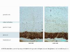
detail of olfactory bulb layers with (left) and without (right) 6-OHDA previously applied to the median forebrain bundle
|
| filename, dt-27-olf-detail-TH
(format, JPEG)
|
| file size, 0.08 MiB |
|
size at 300 dpi, 97mm x 73mm/ 3.84" x 2.88"
|
| date, 20 08 2005
|
| to see the image full-size, click the thumbnail |
|
|
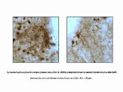
high power view of single glomerulus after 6-OHDA administration to mfb three weeks previously (left). S19 TH immunohistochemistry.
|
| filename, dt-28_glomeruli
(format, JPEG)
|
| file size, 0.05 MiB |
|
size at 300 dpi, 97mm x 73mm/ 3.84" x 2.88"
|
| date, 20 08 2005
|
| to see the image full-size, click the thumbnail |
|


 main index