|
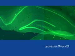
the hippocampus labelled by Neuronal N immunohistochemistry
|
| filename, dt_xOH1_NeuN0505 (format, JPEG) |
| file size, 0.07 MiB |
| size at 300 dpi, 97mm x 73mm/3.84" x 2.88" |
| date, 31 05 2005 |
| to see the image full-size, click the thumbnail |
|
|
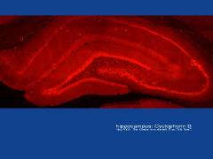
architecture of the hippocampus in coronal section; Cyclophilin B immunohistochemistry
|
| filename, dt_xOH1_CycloPhB0505 (format, JPEG) |
| file size, 0.07 MiB |
|
size at 300 dpi, 97mm x 73mm/ 3.84" x 2.88"
|
| date, 31 05 2005
|
| to see the image full-size, click the thumbnail |
|
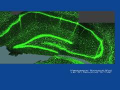
| rat hippocampus labelled with green fluorescent Nissl stain |
| filename, xOH7dtfluoroNissl4 (format, JPEG) |
| file size, 0.14 MiB |
| size at 300 dpi, 97mm x 73mm/3.84" x 2.88" |
| date, 15 08 2005 |
| to see the image full-size, click the thumbnail |
|
|
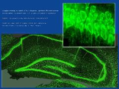
general view and CA1 detail of rat hippocampus labelled with green fluorescent Nissl stain
|
| filename, xOH7dtfluoroNsslComp
(format, JPEG)
|
| file size, 0.13 MiB |
|
size at 300 dpi, 97mm x 73mm/ 3.84" x 2.88"
|
| date, 02 07 2005
|
| to see the image full-size, click the thumbnail |
|
|
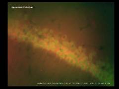
CA1 region of rat hippocampus, imaged with nuclear and cytoplasmic stains
|
| filename, dt_xOH1_CA1_0505
(format, JPEG)
|
| file size, 0.06 MiB |
|
size at 300 dpi, 97mm x 73mm/ 3.84" x 2.88"
|
| date, 31 05 2005
|
| to see the image full-size, click the thumbnail |
|
|
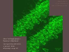
CA1 region of hippocampus (confocal microscope views of adjacent slices), labelled with fluorescent green Nissl stain
|
| filename, xOH7dtConfocalGrnNissl
(format, JPEG)
|
| file size, 0.13 MiB |
|
size at 300 dpi, 97mm x 73mm/ 3.84" x 2.88"
|
| date, 15 08 2005
|
| to see the image full-size, click the thumbnail |
|
|
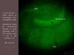
rat hippocampal organotypic slice, labelled for Cyclophilin B
|
| filename, OH2_dt_CycloB_x10
(format, JPEG)
|
| file size, 0.07 MiB |
|
size at 300 dpi, 97mm x 73mm/ 3.84" x 2.88"
|
| date, 15 08 2005
|
| to see the image full-size, click the thumbnail |
|
|
CA1 region of rat hippocampus labelled for Cyclophilin B
|
| filename, OH2_dt_CA2CycloB
(format, JPEG)
|
| file size, 0.12 MiB |
|
size at 300 dpi, 97mm x 73mm/ 3.84" x 2.88"
|
| date, 15 08 2005
|
| to see the image full-size, click the thumbnail |

|
|
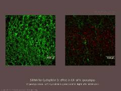
effect of interfering RNA (siRNA) on Cyclophilin B expression (right) in organotypic hippocampal slice
|
| filename, OH3_dt_siRNA_CyBalexa
(format, JPEG)
|
| file size, 0.15 MiB |
|
size at 300 dpi, 97mm x 73mm/ 3.84" x 2.88"
|
| date, 15 08 2005
|
| to see the image full-size, click the thumbnail |
|
|
effect of interfering RNA (siRNA) on Cyclophilin B expression (right) in organotypic hippocampal slice
|
| filename, OH3x40dtsiRNA_CyPhB
(format, JPEG)
|
| file size, 0.11 MiB |
|
size at 300 dpi, 97mm x 73mm/ 3.84" x 2.88"
|
| date, 15 08 2005
|
| to see the image full-size, click the thumbnail |
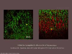
|
|
appearance of siRNA for Cyclophilin B, Cy3 label
|
| filename, 0507OH12dtsiRNA
(format, JPEG)
|
| file size, 0.09 MiB |
|
size at 300 dpi, 97mm x 73mm/ 3.84" x 2.88"
|
| date, 08 08 2005
|
| to see the image full-size, click the thumbnail |
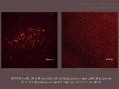
|
|
confirmation of effect of siRNAon Cyclophilin B expression (right). Third label, DAPI for nuclei (blue).
|
| filename, 0507xOH12siRNAcompar
(format, JPEG)
|
| file size, 0.12 MiB |
|
size at 300 dpi, 97mm x 73mm/ 3.84" x 2.88"
|
| date, 01 08 2005
|
| to see the image full-size, click the thumbnail |
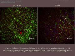
|
|
rat hippocampus labelled with activated glial fibrillary acidic protein (aGFAP), a marker for glia
|
| filename, 0509xOH17dtaGFAPHC
(format, JPEG)
|
| file size, 0.108 MiB |
|
size at 300 dpi, 97mm x 73mm/ 3.84" x 2.88"
|
| date, September 2005
|
| to see the image full-size, click the thumbnail |
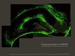
|
|
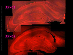
a comparison of hippocampus labelled with two antibodies to TRPV4
|
| filename, 0511XOH18TRPV4cmp
(format, JPEG)
|
| file size, 0.2 MiB |
|
size at 300 dpi, 97mm x 73mm/ 3.84" x 2.88"
|
| date, November 2005
|
| to see the image full-size, click the thumbnail |
|
|
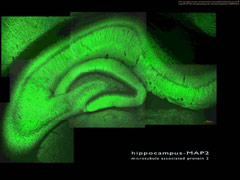
general view of rat hippocampus labelled for microtubule associated protein 2 (MAP2)
|
| filename, f20509xOH17MAP2HC
(format, JPEG)
|
| file size, 0.12 MiB |
|
size at 300 dpi, 97mm x 73mm/ 3.84" x 2.88"
|
| date, September 2005
|
| to see the image full-size, click the thumbnail |
|
|
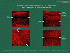
density of GFAP-labelled astrocytes in the pyramidal cell layers of the rat hippocampus |
| filename, 0509xOH17dtGFAPinHC
(format, JPEG)
|
| file size, 0.1 MiB |
|
size at 300 dpi, 97mm x 73mm/ 3.84" x 2.88"
|
| date, September 2005
|
| to see the image full-size, click the thumbnail |
|
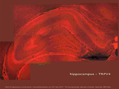
| distribution of TRPV4 in rat hippocampus using immunohistochemical labelling |
| filename, 0509xOH17dtTRPV4HC (format, JPEG) |
| file size, 0.15 MiB |
| size at 300 dpi, 97mm x 73mm/3.84" x 2.88" |
| date, September 2005 |
| to see the image full-size, click the thumbnail |
|
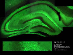
| immunohistochemical distribution of synapsin in the rat hippocampus |
| filename, 0511xOH19synapsinMsc (format, JPEG) |
| file size, 0.1 MiB |
| size at 300 dpi, 97mm x 73mm/3.84" x 2.88" |
| date, November 2005 |
| to see the image full-size, click the thumbnail |
|


 main index