|
overall view of rat brain, in parasagittal section, using myelin (Luxol fast blue) and neuronal (Nissl) stains
|
| filename, dt_23_rat_parasagittal
(format, JPEG)
|
| file size, 0.09 MiB |
|
size at 300 dpi, 97mm x 73mm/ 3.84" x 2.89"
|
| date, 20 08 2005
|
| to see the image full-size, click the thumbnail |

|
|
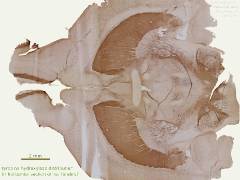
tyrosine hydroxylase labelling of rat forebrain in horizontal section, with, from left, glomerular layers of OB, striatum, and Substantia Nigra
|
| filename, dt_29_panTH_composite
(format, JPEG)
|
| file size, 0.15 MiB |
|
size at 300 dpi, 97mm x 73mm/ 3.84" x 2.88"
|
| date, 20 08 2005
|
| to see the image full-size, click the thumbnail |
|
|
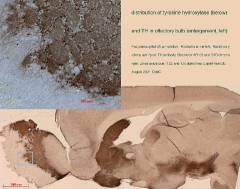
summary figure showing tyrosine hydroxylase anatomy of the rat brain (below), and detail of the glomeruli of the olfactory bulb (top left)
|
| filename, dt_panTH-6OHDA-mosaic
(format, JPEG)
|
| file size, 0.11 MiB |
|
size at 300 dpi, 97mm x 77mm/ 3.84" x 3.03"
|
| date, 20 08 2005
|
| to see the image full-size, click the thumbnail |
|
|
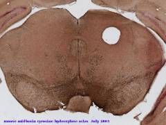
slide from reference series of tyrosine hydroxylase labelled coronal sections prepared from mouse hindbrain
|
| filename, desktopTHatlas
(format, JPEG)
|
| file size, 0.11 MiB |
|
size at 300 dpi, 97mm x 73mm/ 3.84" x 2.88"
|
| date, 20 08 2005
|
| to see the image full-size, click the thumbnail |
|
|
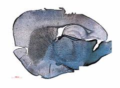
brain of mouse in parasagittal section, showing tyrosine hydroxylase (brown) in SN, STN and striatum. Blue, myelin.
|
| filename, dt_striatum_composite
(format, JPEG)
|
| file size, 0.09 MiB |
|
size at 300 dpi, 97mm x 72mm/ 3.84" x 2.83"
|
| date, 20 08 2005
|
| to see the image full-size, click the thumbnail |
|
|
detail from previous figure showing striatum and thalamus of mouse, showing tyrosine hydroxylase (brown) in striatum. Blue, myelin.
|
| filename, dt_striatum
(format, JPEG)
|
| file size, 0.11 MiB |
|
size at 300 dpi, 97mm x 77mm/ 3.84" x 3.04"
|
| date, 20 08 2005
|
| to see the image full-size, click the thumbnail |
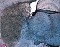
|
|
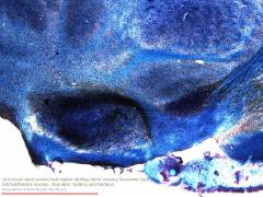
dark masses of the Substantia Nigra (lower centre) and the Subthalamic Nucleus (centre right) in the ventral midbrain, TH and LFB labels
|
| filename, dt_midbrain_panTH
(format, JPEG)
|
| file size, 0.09 MiB |
|
size at 300 dpi, 97mm x 73mm/ 3.84" x 2.88"
|
| date, 20 08 2005
|
| to see the image full-size, click the thumbnail |
|
|
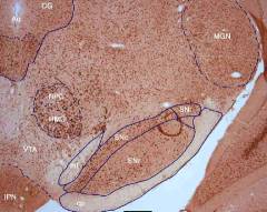
regions of ventrolateral rat midbrain in transverse section labelled immunohistochemically for Hu. Image by Tharushini Bowala.
|
| filename, Hu_labeling_in_rat_mid
(format, JPEG)
|
| file size, 0.09 MiB |
|
size at 300 dpi, 97mm x 77mm/ 3.84" x 3.06"
|
| date, 20 08 2005
|
| to see the image full-size, click the thumbnail |
|
|
early experiment showing effect of previous unilateral 6-OHDA injection in striatum on the ipsilateral Substantia Nigra (top left)
|
| filename, desktopTHmontage
(format, JPEG)
|
| file size, 0.07 MiB |
|
size at 300 dpi, 97mm x 73mm/ 3.84" x 2.88"
|
| date, 20 08 2005
|
| to see the image full-size, click the thumbnail |
|
|
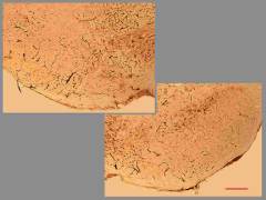
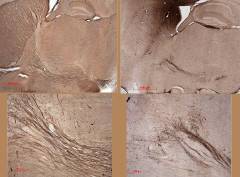
the nigrostriatal tract, enlarged below, showing on the right the toxic effects of 6-OHDA administered within the striatum
|
| filename, dt_fibres
(format, JPEG)
|
| file size, 0.13 MiB |
|
size at 300 dpi, 97mm x 72mm/ 3.84" x 2.83"
|
| date, 15 08 2005
|
| to see the image full-size, click the thumbnail |
|
|
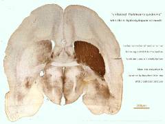
horizontal section of rat forebrain showing deficit of striatal tyrosine hydroxylase after ipsilateral midbrain forebrain bundle 6-OHDA, in model of PD
|
| filename, dt-27-6OHDA-forebrain
(format, JPEG)
|
| file size, 0.1 MiB |
|
size at 300 dpi, 97mm x 73mm/ 3.84" x 2.88"
|
| date, 20 08 2005
|
| to see the image full-size, click the thumbnail |
|
|
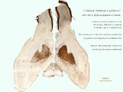
horizontal section rat forebrain labelled for tyrosine hydroxylase after ipsilateral midbrain forebrain bundle 6-OHDA. Note effect on striatum but not OB
|
| filename, dt-27-6OHDAuniOlfact
(format, JPEG)
|
| file size, 0.1 MiB |
|
size at 300 dpi, 97mm x 73mm/ 3.84" x 2.88"
|
| date, 20 08 2005
|
| to see the image full-size, click the thumbnail |
|
|
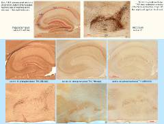
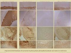
tyrosine hydroxylase phosphorylation variants: distribution in the rat hippocampus and midbrain. Parasagittal sections.
|
| filename, TH-comparison_peroxidase
(format, JPEG)
|
| file size, 0.12 MiB |
|
size at 300 dpi, 97mm x 73mm/ 3.84" x 2.9"
|
| date, 20 08 2005
|
| to see the image full-size, click the thumbnail |
|
|
distribution of tyrosine hydroxylase phosphorylation variants in rat olfactory bulb, striatum, Substantia Nigra and adrenal gland
|
| filename, dt_29_TH_comparison
(format, JPEG)
|
| file size, 0.12 MiB |
|
size at 300 dpi, 97mm x 73mm/ 3.84" x 2.88"
|
| date, 20 08 2005
|
| to see the image full-size, click the thumbnail |
|
|
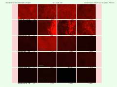
studies of tyrosine hydroxylase phosphorylation variants in the rat striatum
|
| filename, desktop_TH_comparison
(format, JPEG)
|
| file size, 0.12 MiB |
|
size at 300 dpi, 97mm x 73mm/ 3.84" x 2.88"
|
| date, 20 08 2005
|
| to see the image full-size, click the thumbnail |
|
|
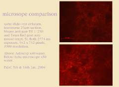
comparison of fluorescent image from two microscopes
|
| filename, desktopmicroscope
(format, JPEG)
|
| file size, 0.12 MiB |
|
size at 300 dpi, 97mm x 71mm/ 3.84" x 2.8"
|
| date, 20 08 2005
|
| to see the image full-size, click the thumbnail |
|


 main index