

Reading the Volumes
(In the pulmonary function laboratory)
Reference ranges.
Hemoglobin, adult, g/l. Female 120-156, male 138-172.
Carboxyhemoglobin. Less than 2%.
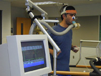
Mrs A, Mrs B and Dr C are in the pulmonary function testing room. Mrs A, aet. 48, has scoliosis, a rotational deformity of the spine. She is planning to have an orthopedic procedure requiring general anesthesia, and the anesthetist has requested evaluation of her pulmonary function. Mrs B, 38, has been referred for investigation of gradually increasing shortness of breath with exertion. Dr C, 28, has volunteered to act as a normal control. Each of them is well in other ways and none of them smokes.
Weights and heights are, respectively (kg:m): Mrs A, 58:1.7; Mrs B, 82:1.65; Dr C, 72:1.7.
* Calculate their BMI (kg/m2).
Mrs A, 20; Mrs B, 30; Dr C, 25.
* Could these have any significance in the present setting?
Obesity may restrict lung expansion. Low body weight, on the other hand, may indicate increased respiratory work.
Results from blood tests arranged the previous day are as follows. Hemoglobin (g/l) Mrs A, 145; Mrs B, 155; Dr C, 135.
* Suggest interpretations of these values from a respiratory perspective.
A raised Hb (polycythemia-here, borderline) is likely to be a response to chronic hypoxemia.
Carboxyhemoglobin (percentage of hemoglobin): Mrs A, undetectable; Mrs B, 12; Dr C, 4.
* What does COHb indicate?
Carboxyhemoglobin is hemoglobin bound by carbon monoxide.
* Suggest interpretations of these values.
Mrs B smokes heavily and Dr C admits that she lives with smokers.
Peak flow testing for Dr C gives 320, 410 and 420 l/min.
* What are advantages and disadvantages of this test?
The apparatus is cheap and widely available. The test can be carried out easily and by staff with brief training. A disadvantage is that results are very reliant on subject effort, so it is more useful for comparisons over time in an individual than for comparing individuals.

Using a spirometer, forced expiratory volume in 1 s. (FEV1) and forced vital capacity (FVC) are measured with the following results (shown above). FEV1 (litres) Mrs A, 2.7; Mrs B, 1; Dr C, 4. FVC (l) Mrs A, 3; Mrs B, 3; Dr C, 5.
* Calculate the FEV1/FVC ratio for each person and comment on the findings.
Mrs A, 0.9; Mrs B, 0.33; Dr C, 0.8. The ratio is above normal for Mrs A, but the significant finding in her case is the reduced vital capacity. Mrs B also has a reduced vital capacity, but her FEV1 is markedly reduced. Dr C has a normal FEV1 and FVC.
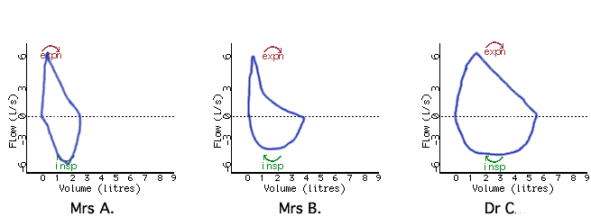
Using a pneumotachograph, rate of airflow is measured during quiet breathing and gas volumes are calculated. The subjects are asked to exhale forcefully from maximum inspiration, then to inhale again to vital capacity, which is the origin in these recordings. The plotted flow/volume loops are as follows.

* Which points on these loops represent Vital Capacity and Residual Volume?
Vital Capacity is the point at the far left of each loop, where flow is zero and inspiratory volume is maximal. Residual Volume is the point at the far right: flow is also zero, and expiration is complete.
* Could Peak Flow and Vital Capacity be obtained from these flow/volume curves?
Yes: Peak Flow is the greatest rate of expiration, simply read from the 'flow' axis as the highest point reached on the expiratory curve, and Vital Capacity can be quantified as the difference along the "volume" axis between the point where Vital Capacity is reached (see previous question) and the 'residual volume' point.
The expiratory part of the loop has an asymmetrical course, rising quickly to a high flow rate and then falling slowly.
* Is airway resistance to airflow greater in inspiration or in expiration?
Airway resistance is greater in expiration (Einthoven, 1892), because during expiration the pressure in the lung surrounding the airways is higher than in the lumen, compressing the lumen. In inspiration, the surrounding parenchyma is at a lower pressure than inside the lumen, causing it to widen.
* Why are there two phases in the expiratory flow trajectories?
In the first part of expiration air is expelled from low-resistance large airways. In the subsequent part there is a balance between airway collapse and expiratory flow.
* Why is this phenomenon more marked in the recording from Mrs B?
Deterioration of elastic tissue around alveoli results in less support for small airways during expiration, allowing them to collapse more easily.
* Can you suggest a diagnosis for Mrs B's pulmonary condition?
She is likely to have chronic obstructive pulmonary disease (COPD/CORD etc). The emphysematous type causes particular reduction in expiratory flow at low rates and lengthening of forced expiratory duration.
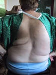
| * Can you interpret the curve for Mrs A in terms of her condition? The flow-volume loop shows that flow is not reduced, but lung volume is markedly restricted. Aspects of chest wall anatomy including movement of the ribs at their articulation on the spine are important for lung volume change during ventilation. In scoliosis, the asymmetrical movement of the ribs may limit the extent of lung expansion. If ventilation is insufficient for oxygen transfer she will be dyspneic; this is typically worse with exercise, when oxygen demand increases. |

Moderate scoliosis picture: Richard & Debra Austin |
Details about the metacholine challenge test from Medical Centre East, Birmingham Alabama.
A good general review of respiratory physiology can be got by browsing through the 24 brief items in the Johns Hopkins School of Medicine's Interactive Respiratory Physiology Encyclopedia.
Corrections and suggestions to D de Castro please.
(problem based on Guenter CA et al. Clinical Aspects of Respiratory Physiology
Lippincott '78 & CIBA Clinical Symposia 1979 Annual with illustrations by Frank Netter)