figure adapted from Goldberg, M, Green, SB, Moss, ML et al. 1973 JAMA 223:269


A 19 year old male is brought to a doctor's office in Khandallah by his mother
who is very worried. He was found shortly earlier in considerable distress
complaining that he was unable to breathe. He is unable to provide a coherent
history.
On examination his respiratory rate is markedly increased at rest to nearly 50/min at times. There is no cough. There is no cyanosis (blue tint of lips suggesting a high level of de-oxygenated hemoglobin ) or pallor (reduction in the red colour of the conjunctival mucosa suggesting anemia). He is diaphoretic (sweaty) but afebrile. He is able to speak in short sentences but seems anxious and only answers simple questions. He is making use of accessory respiratory muscles and his breaths are deep with marked chest wall movement. On auscultation of his chest there are prominent air sounds with no adventitiae (added sounds).
* what is a normal adult resting respiratory rate (from your own experience)?
For discussion; rather variable--perhaps around 12/min
* is this asthma, or an airway problem generally?
In asthma there is airway obstruction (due to bronchospasm): this patient
is moving large amounts of air as indicated by the large chest wall movement.
The narrowed airways in asthma also produce abnormal, 'musical' sounds (rhonchi)
on auscultation, which are not present here. There are 'prominent air sounds'—i.e. sounds of normal air movement—on chest auscultation.
Note however that bronchospasm is a possible complication of this condition, in those with 'sensitivity' of the airway surfaces. Voluntary hyperventilation is used as a test for bronchial hypersensitivity/asthma as an alternative to inhaled methacholine.
Thus this does not seem to be an 'airway' problem.
* what will be the effect of increased ventilation on the partial pressure of carbon dioxide in the arterial blood-an increased level (hypercapnia) or a decreased level (hypocapnia)?
Hypocapnia: PaC02 is reduced.

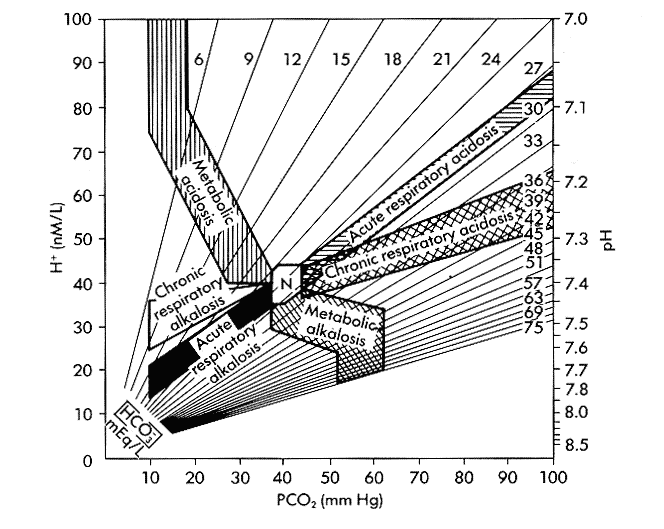
figure adapted from Goldberg, M, Green, SB, Moss, ML et al. 1973 JAMA 223:269
The diagram above is one way of laying out in a plane the three variables
involved in acid-base balance in the blood (PaCO2-the partial pressure of
arterial CO2, HCO3--the concentration of the bicarbonate ion, and pH, the
degree of acidity or alkalinity of the blood). The pH can also be expressed
as [H+], the concentration of hydrogen ions (protons). It shows that given
any two of these three values the third is determined. Note that oxygen
carriage is not directly related to acid-base status. This 'nomogram' is
used for the interpretation of the results of arterial blood gas tests. The
region labelled 'N' indicates normal acid-base values.
* If there is little change in [HC03] so far in this patient, what is the
effect of the change in PaCO2 noted in the last question on the pH of the
blood?
The pH will be increased.
* What is the category of acid-base disturbance exhibited by this patient?
"Acute respiratory alkalosis"
For an interactive, and fun, version of this nomogram see this site at the University of Tulane Department of Anesthesia. Note that the axes are arranged somewhat differently here from those in the nomogram above.
The patient says between gasps that he has cramping in the arms and tingling
and numbness.
* an increase in blood pH (alkalosis) has some general (systemic) effects
on the body, particularly on the musculoskeletal and nervous systems. Can
you suggest ways in which this might occur?
Alkalosis may have systemic effects by various mechanisms, including
- lowered pH of cerebrospinal fluid (CSF) causing reflex cerebral vasoconstriction
and hypoxia
- decreased delivery of oxygen to tissues due to increase in oxygen binding
to hemoglobin at low PO2 (the "Bohr effect")
- increased binding of calcium ions in blood to carrier proteins causing
effective hypocalcemia. Calcium is largely bound to albumin in the blood
and at increased pH more calcium is bound, reducing the ionised fraction
and causing a relative hypocalcemia. Calcium is important for nerve and
muscle function.
Calcium (Ca) is required for the proper functioning of numerous intracellular
and extracellular processes, including muscle contraction, nerve conduction, hormone release and blood coagulation. In addition, the Ca ion plays a unique
role in intracellular signaling and is involved in the regulation of many
enzymes. The maintenance of Ca homeostasis, therefore, is critical. (Merck
Manual of Therapeutics, ch. 12)
Hypocalcemia causes the clinical syndrome of 'tetany'.
Tetany characteristically results from severe hypocalcemia. It can also result
from reduction in the ionized fraction of plasma Ca without marked hypocalcemia,
as occurs in severe alkalosis. Tetany is characterized by sensory symptoms
consisting of paresthesias of the lips, tongue, fingers and feet; carpopedal
spasm, which may be prolonged and painful; generalized muscle aching; and
spasm of facial musculature. Tetany may be overt with spontaneous symptoms
or latent and requiring provocative tests to elicit. Latent tetany generally
occurs at less severely decreased plasma Ca concentrations: 7 to 8 mg/dL
(1.75 to 2.20 mmol/L). (Merck Manual of Therapeutics, ch. 12)
It is not clear how these effects of alkalosis combine in the brain to maintain
the abnormal signal to the lungs ('increased ventilatory drive').
Note: One physiological function which is likely to be important in the phenomenon of 'tetany' is the requirement for calcium ions in the process of neurotransmitter release at the axon terminals of nerves.
Instructing the patient to reduce the rate and depth of breathing has no
effect. A feedback loop is occuring, which is maintaining ventilation at
abnormal values.
* a similar feedback loop can maintain disordered function in other systems.
Can you give an example?
Singultus (hiccups)
* bearing in mind that ventilation is controlled from the caudal brainstem (medulla oblongata) rather than by the lungs themselves, can you sketch a figure indicating the course of this circuit?
'Respiratory centre' in medulla oblongata (--> phrenic nerves etc) --> diaphragm
and other muscles of ventilation --> hypocapnia --> alkalosis --> hypocalcemia
and other metabolic derangements --> altered cerebral function with "anxiety" ?limbic system
activation --> 'respiratory centre' in medulla oblongata -->
The young doctor does not recognise the syndrome and sends him off to a hospital.
However, in the ambulance the experienced paramedics resolve his respiratory
distress with a simple manouvre.
* what is the manouvre?
Having the patient re-breathe his expired air from a paper bag
* how does it solve the problem?
Expired air contains more CO2 than atmospheric air. An increased level of
inhaled C02 (FICO2), increases alveolar CO2 (PAC02), increases arterial CO2
(PaCO2), and reverses systemic alkalosis. Plot this correction on the nomogram
above.
* can you suggest other causes of "hyperventilation" besides the syndrome
described here?
Many important conditions can cause increased respiratory efforts leading
to hypocapnia. These include conditions acting in the lung (pulmonary embolism-blockage
of the pulmonary blood vessels; pneumonia-lobar consolidation of lung-etc),
in the brain (example, salicylate poisoning), or in other systems (e.g. heart
failure, severe acute blood loss).
"Hyperventilation" refers to a situation in which hypocapnia results. Do not use the word
carelessly to mean only the phenomenon ("Hyperventilation Syndrome") described in this case study.
The mother quietly informs the doctor that there was a possible precipitating
event involving a break-up with a girlfriend two days before.
* in what proportion of cases does the doctor ever find out what really went on?
References
John Laffey and Brian Kavanagh: Hypocapnia. N Engl J Med. 2002 Jul 4;347(1):43-53.
(local copy here)
Stephen Gluck: Acid-base. Lancet 1998; 352: 47479 (local copy
here). Respiratory acid-base disorders are mentioned in the final
paragraph. Corrections and suggestions to D de Castro please.
(problem based on case seen by DdeC about 1985)